Cellular Illustration (Cell Structure and Function)
The Cellular Illustration portfolio showcases the intricate structures and mechanisms of different cells.
Click on each illustration below to enlarge.
Cellular Illustration: Stages of Mitosis - This illustration about the stages of mitosis was created for medical education. Mitosis is a type of cell division that happens in most of the cells in the body. One cell divides to create two daughter cells that are genetically identical to the original cell. During mitosis, the replicated chromosomes align, and split into two complete sets for each daughter cell.
Cellular Illustration: Centromere Vocabulary - The centromere is located between the two sister chromatids, holding them together. The kinetochore attaches the centromeres to the microtubules before cell division. Together, they power the movement of the chromosomes and separation into sister chromatids.
Cellular Illustration: The Cell Cycle - This illustration shows the entire cell cycle, which can be divided into two main parts: interphase and mitosis. Most of the time, a cell is in interphase, where DNA replication and cell growth occur. Interphase can be divided further into G1 phase, S phase, and G2 phase. G1 and G2 are considered the gap phases. If the conditions around the cell aren't favorable for division, it may enter the G0 phase, where it could remain indefinitely or until the environment is more favorable. The S phase is when DNA replication occurs. After G2, the cell leaves interphase and enters mitosis, where the daughter chromosomes separate and usually end with the cells dividing.
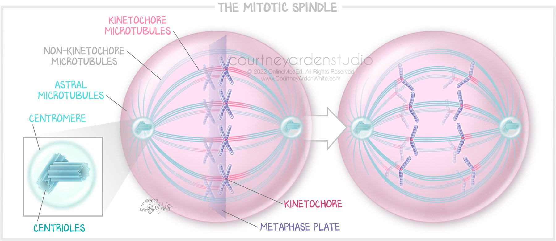
Cellular Illustration: The Mitotic Spindle - This illustration about the mitotic spindle was created for medical education. The mitotic spindle is an essential component of mitosis. It’s composed mostly of microtubules, and its job is to equally divide the chromosomes in the parent cell during cell division.
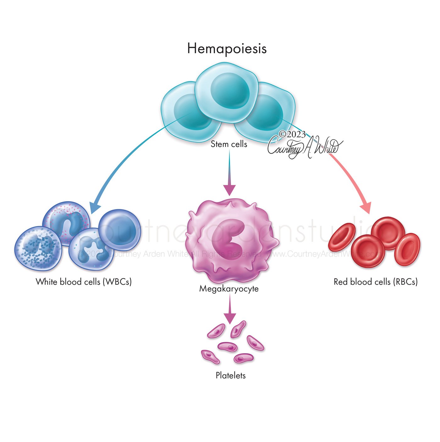
Cellular Illustration: Hemapoiesis
Cellular Illustration: Hepatitis C virus (HCV) assembly
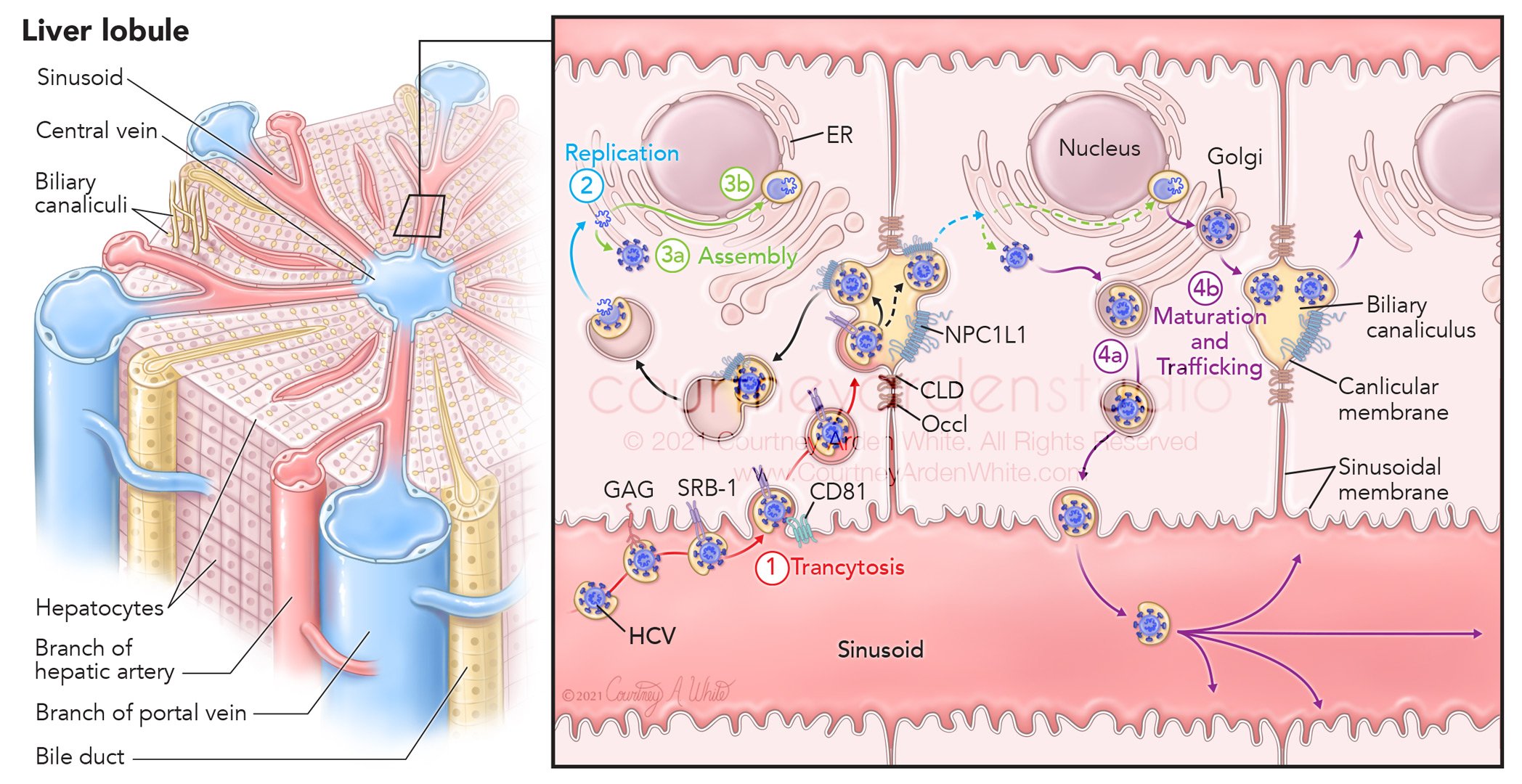
Cellular Illustration: Hepatitis C virus (HCV) replication and assembly and liver lobule anatomy
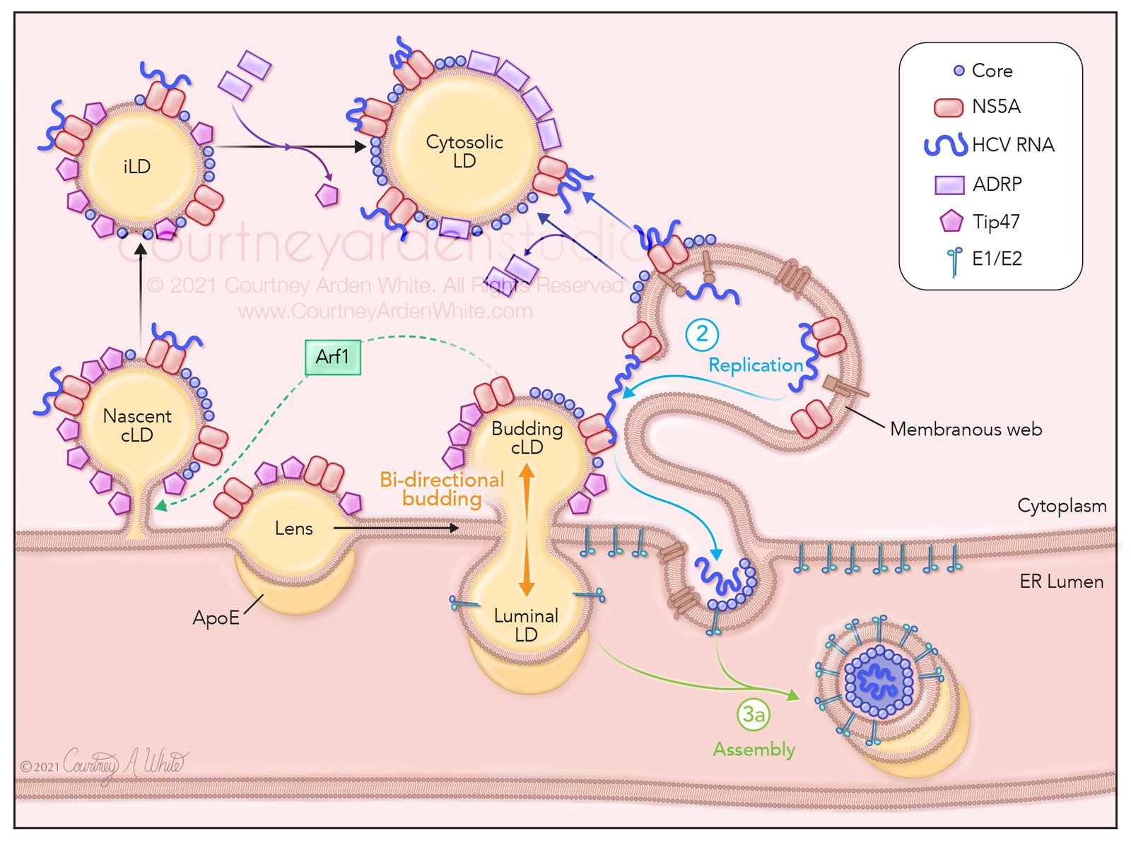
Cellular Illustration: Hepatitis C virus (HCV) budding in ER and cytoplasm


