
The Integumentary System (Skin Illustrations)
The Integumentary System (Skin Illustrations) portfolio includes illustrations of the cells and layers of the skin, including various conditions.
The integumentary system is the body's outer layer and includes the skin (epidermis, dermis, hypodermis), glands, hair, and nails. Its primary function is to act as a protective barrier against harmful external elements and germs while regulating body temperature. The skin is susceptible to various conditions and disorders, as seen in the skin illustrations below, which include psoriasis, skin lesions, skin cancer, eczema, and cellulitis.
Click on each illustration below to enlarge.

Skin Illustrations: Melanocytes - This illustration was created for medical education to explain the role of melanocytes in our body. Melanocytes are cells that produce the pigment melanin. Melanosomes transfer melanin to the surrounding keratinocytes. The melanin then organizes above the nucleus inside each cell and protects the skin from UV radiation. Additionally, melanocytes create pigmentation in the skin and hair. Although everyone has a similar number of melanocytes, the amount of melanin produced varies, resulting in different skin colors. When exposed to sunlight, melanin production increases, giving the appearance of a tan. Keratinocytes degrade the melanin more efficiently in light skin than in dark skin, which is what is shown on the right side of the illustration.

Skin Illustrations: Bruised Thumb

Skin Illustrations: Migrating Epidermal Stem Cells

Skin Illustrations: Injury and Phase 0
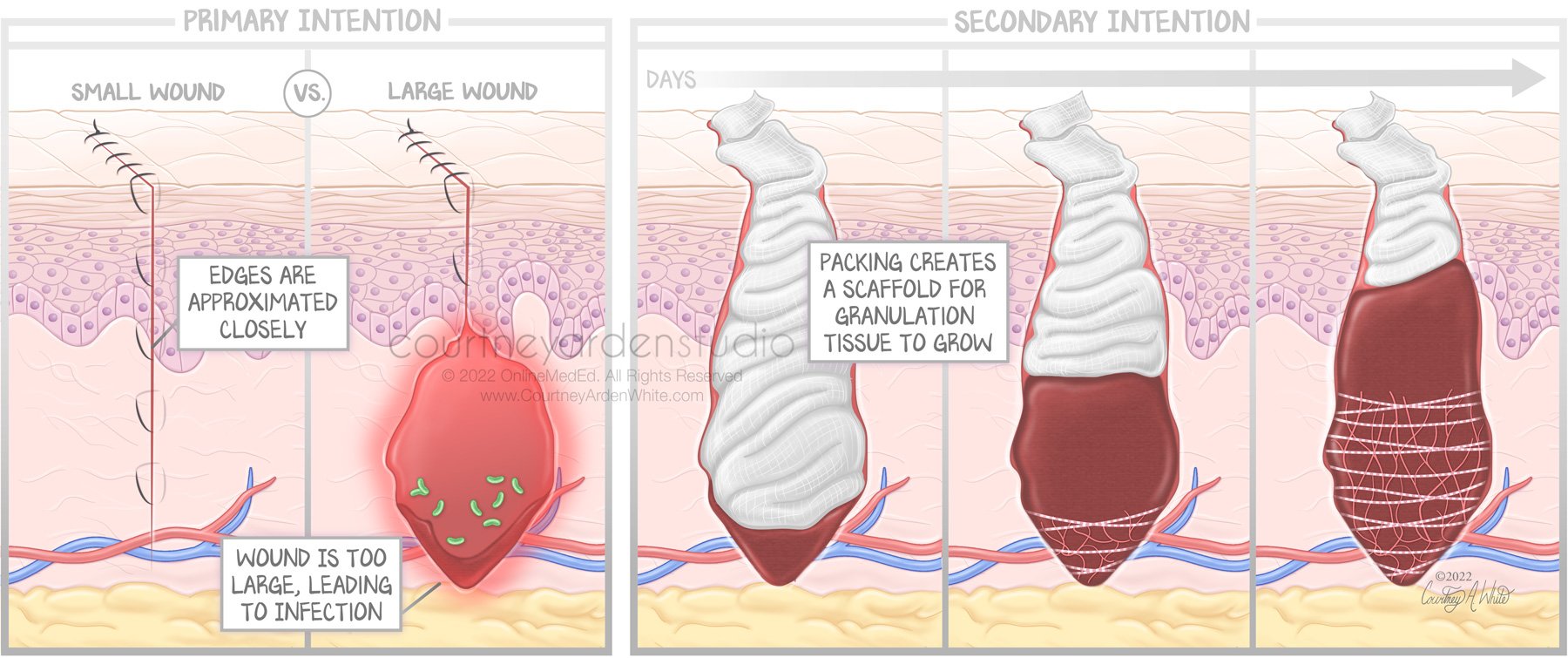
Skin Illustrations: Primary vs. Secondary Intention

Skin Illustrations: Psoriasis Pathogenesis

Skin Illustrations: Nail Root

Skin Illustrations: Pilosebaceous Canal

Skin Illustrations: Glands - Coiled and Straight

Skin Illustrations: Structures of the Hair Shaft

Skin Illustrations: Climate Influences on Prevalence of Childhood Eczema
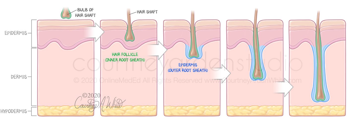
Skin Illustrations: Hair Anatomy - Sinking and Invagination

Skin Illustrations: Appendages are Extensions of the Epidermis

Skin Illustrations: Treatment of Blisters Caused by Running

Skin Illustrations: Erysipelas

Skin Illustrations: Depth of Invasion to Diagnosis
Skin Illustrations: Visualizing the Epidermis

Skin Illustrations: Cells of the Epidermis - Melanocytes
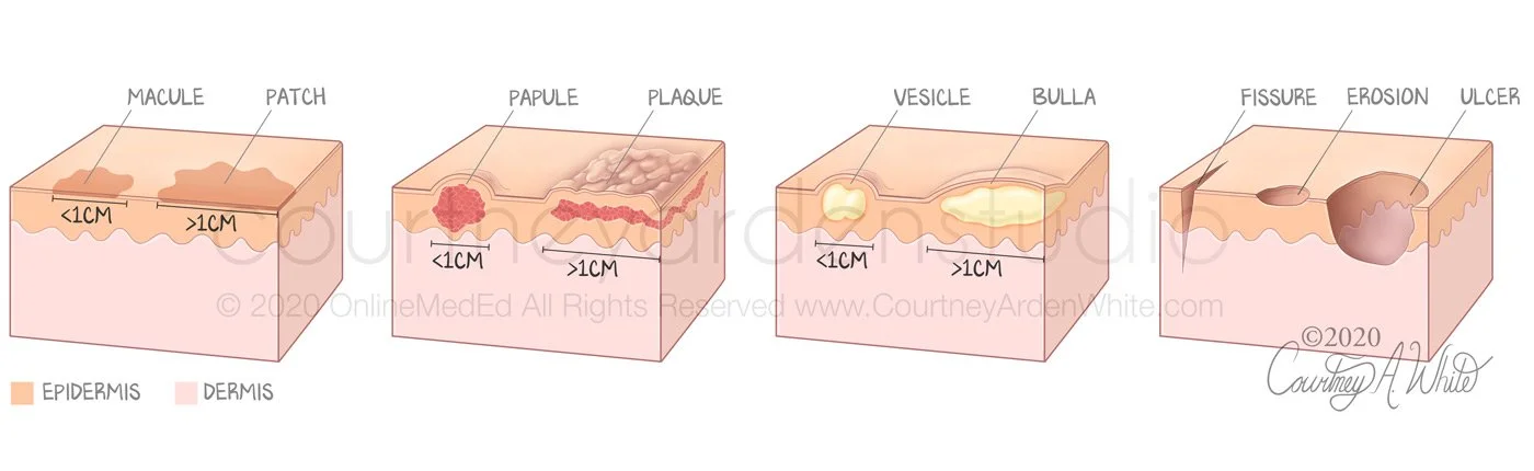
Skin Illustrations: Skin Lesions
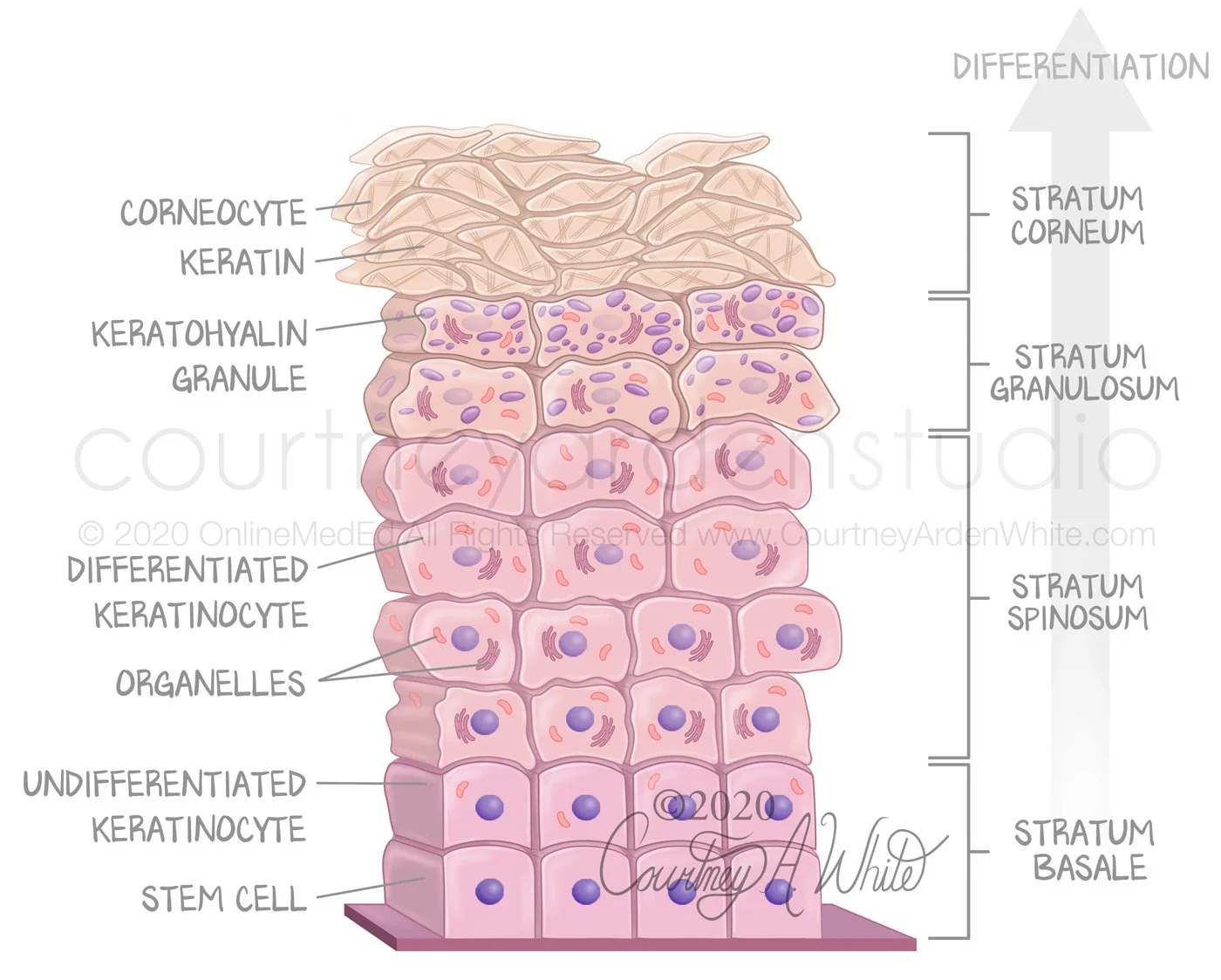
Skin Illustrations: Layers and Cells of the Epidermis

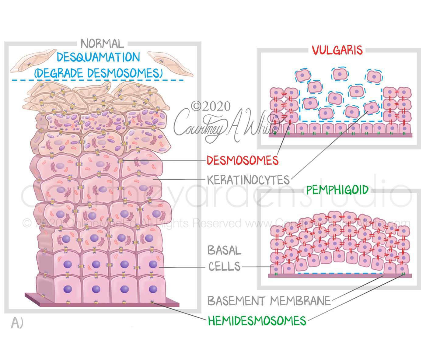
Skin Illustrations: Desmosomes and Proteolytic Activity Leading to Thicker Skin

Skin Illustrations: Layers of the Skin

Skin Illustrations: Skin Cancer Model - This illustration was created for medical education about the histological characteristics of skin cancer as seen under the microscope. The three major types are squamous cell carcinoma, basal cell carcinoma, and Melanoma. Squamous cell carcinoma is composed of spindle cells in a whorled pattern. Basal cell carcinoma is characterized by basaloid nests that stain more blue. Melanoma appears as nested melanocytes of varying sizes.

Skin Illustrations: Skin Fungi