
Urology, Reproduction, and Gynecology Illustration
This Urology, Reproduction, and Gynecology illustration portfolio includes medical illustrations of male and female reproductive and urinary tracts, including healthy anatomy and conditions that can occur in these anatomical areas.
Click on each illustration below to enlarge.
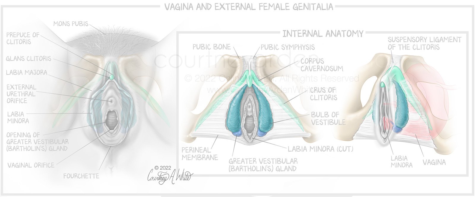
Urology, Reproduction, and Gynecology Illustration: Vagina and External Female Genitalia

Urology Reproduction and Gynecology Illustration: Female-Male Comparison (Clitoris = Penis)
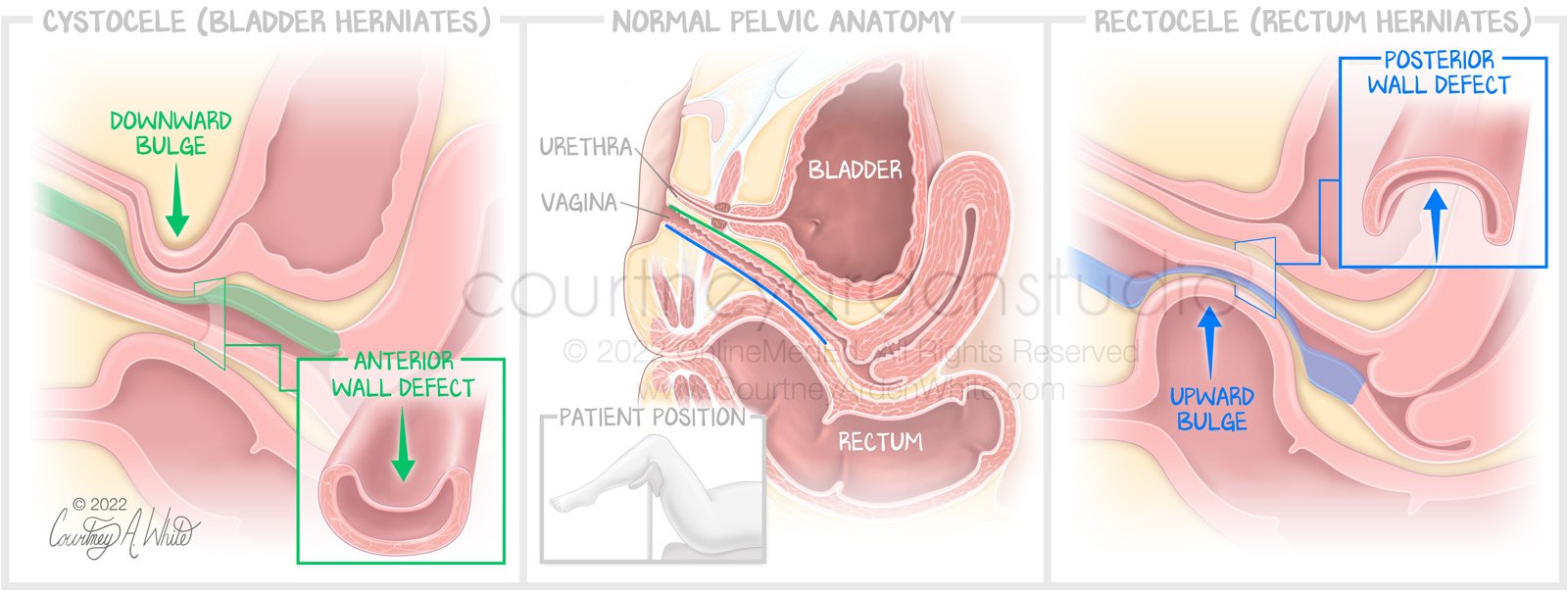
Urology and Reproduction and Gynecology Illustration: Normal Pelvic Anatomy compared to Cystocele (Bladder Herniates) and Rectocele (Rectum Herniates)
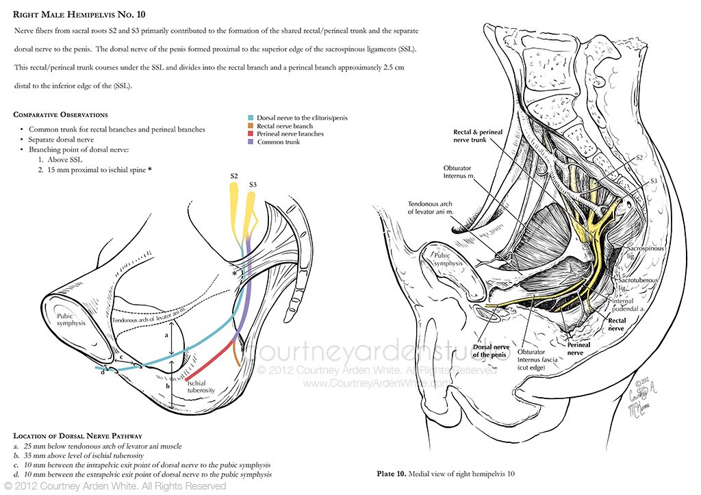
Urology, Reproduction, and Gynecology Illustration: Pudendal Nerve Entrapment Study, hemipelvis #10. This illustration was created as part of my thesis, Pudendal Nerve Entrapment: An Anatomical Study And Three-Dimensional Visualization Of Nerve Variations And Branching Patterns. It is one of 10 hemipelvis dissections I illustrated to study the varied course of the pudendal nerve. Please view the brain and nervous system portfolio for more illustrations from my thesis.
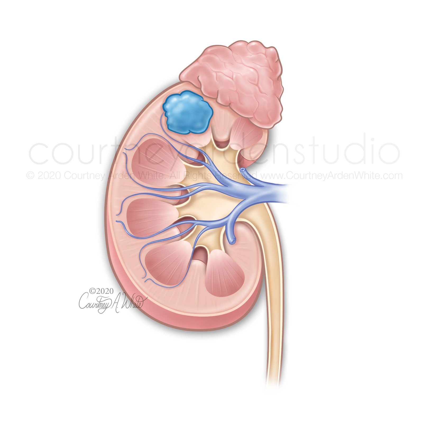
Urology, Reproduction, and Gynecology illustration: This medical illustration showing a tumor in the kidney was created for a two-page patient education handout about kidney cancer. I usually work with other designers and writers, but for this project, I wrote, designed, and illustrated the entire piece. When creating illustrations for patients, it’s important to keep the colors softer, omit anatomical structures that don’t help tell the intended story, and avoid showing blood if it isn’t necessary.
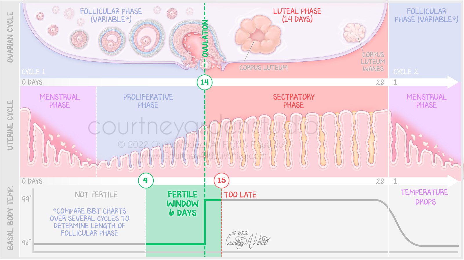
Urology, Reproduction and Gynecology Illustration: Ovarian Cycle, Uterine Cycle and Body Temperature
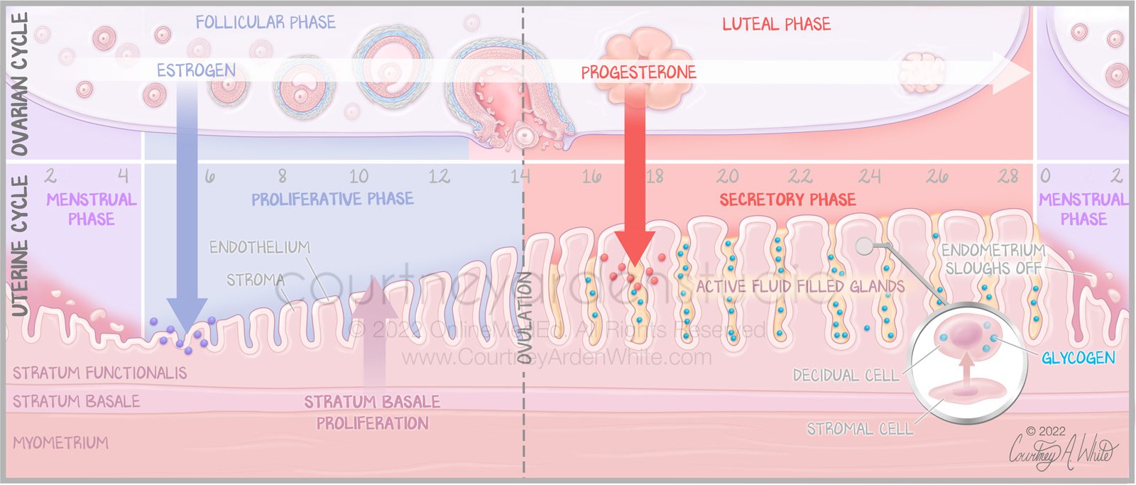
Urology, Reproduction and Gynecology Illustration: Ovarian Cycle, Uterine Cycle, and Uterine wall

Urology, Reproduction, and Gynecology Illustration: Structures of the Renal Corpuscle of the Kidney - The renal corpuscle contains the glomerulus, a group of tiny blood vessels that begin filtration of waste products from the blood and eventually form urine. There are about 1 million renal corpuscles in each kidney.

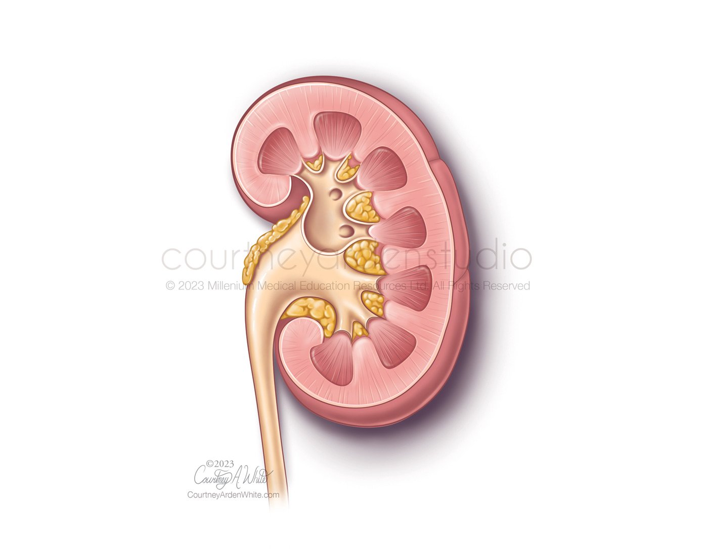
Urology, Reproduction, and Gynecology Illustration: Here is another kidney illustration, but in a different style to match the existing style the client had already created. This illustration provides an anatomical overview of the kidney for medical education. The kidneys' primary function is to filter blood.

Urology Reproduction and Gynecology Illustration: natomy of the bladder, prostate and rectum
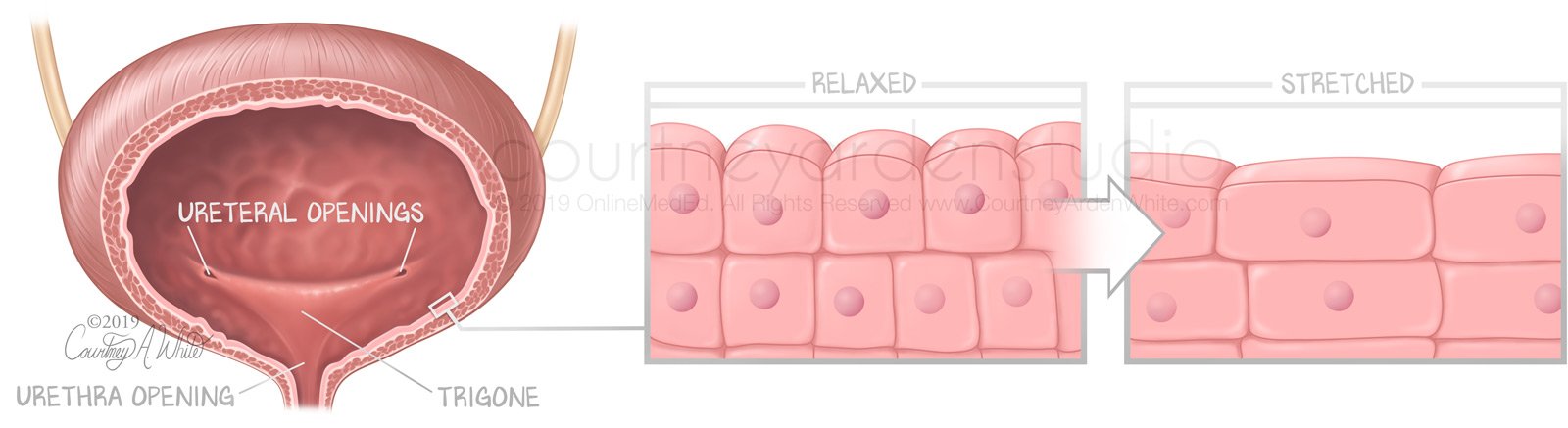
Urology, Reproduction, and Gynecology Illustration: Cells of the bladder wall: This illustration depicts bladder anatomy and shows how the cells relax and stretch when the bladder is empty or full. They were created for medical education for medical students.
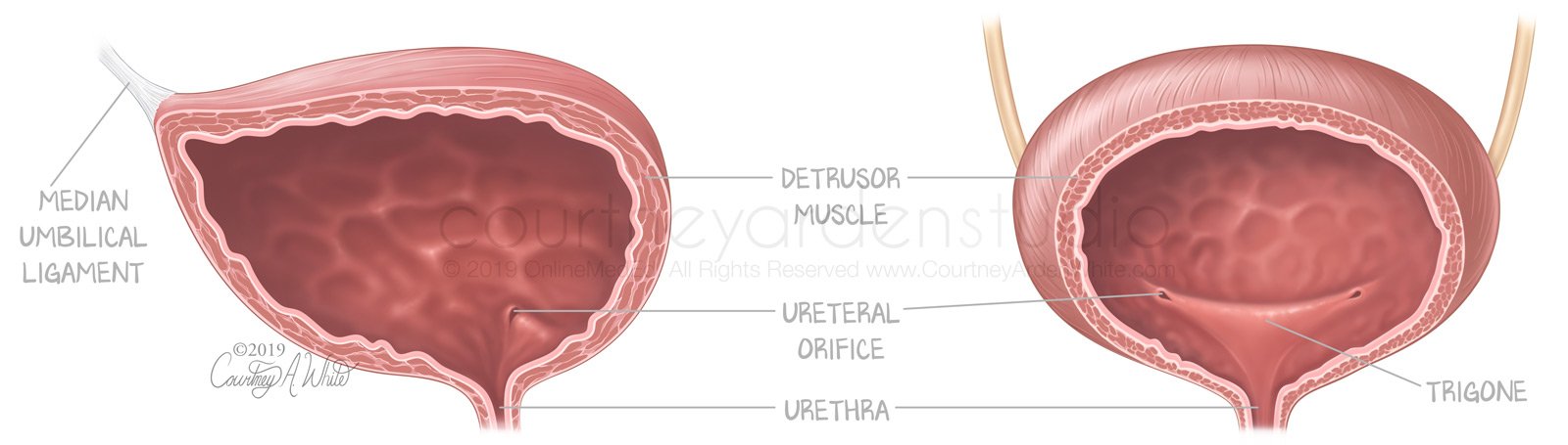
Urology, Reproduction, and Gynecology Illustration: Anatomy of the bladder

Urology, Reproduction Gynecology Illustration: Intracavernosal Injection
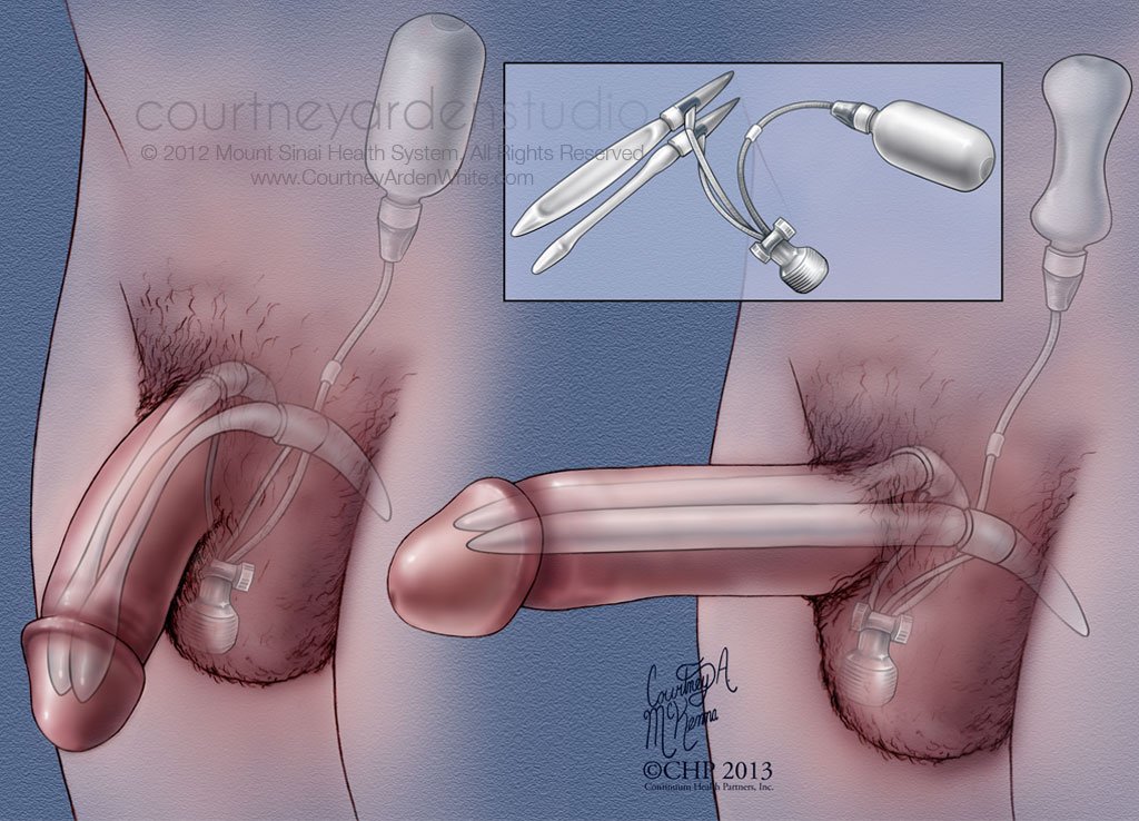
Urology, Reproduction and Gynecology Illustration: penile implant

Urology Reproduction and Gynecology Illustration: kidney nephron
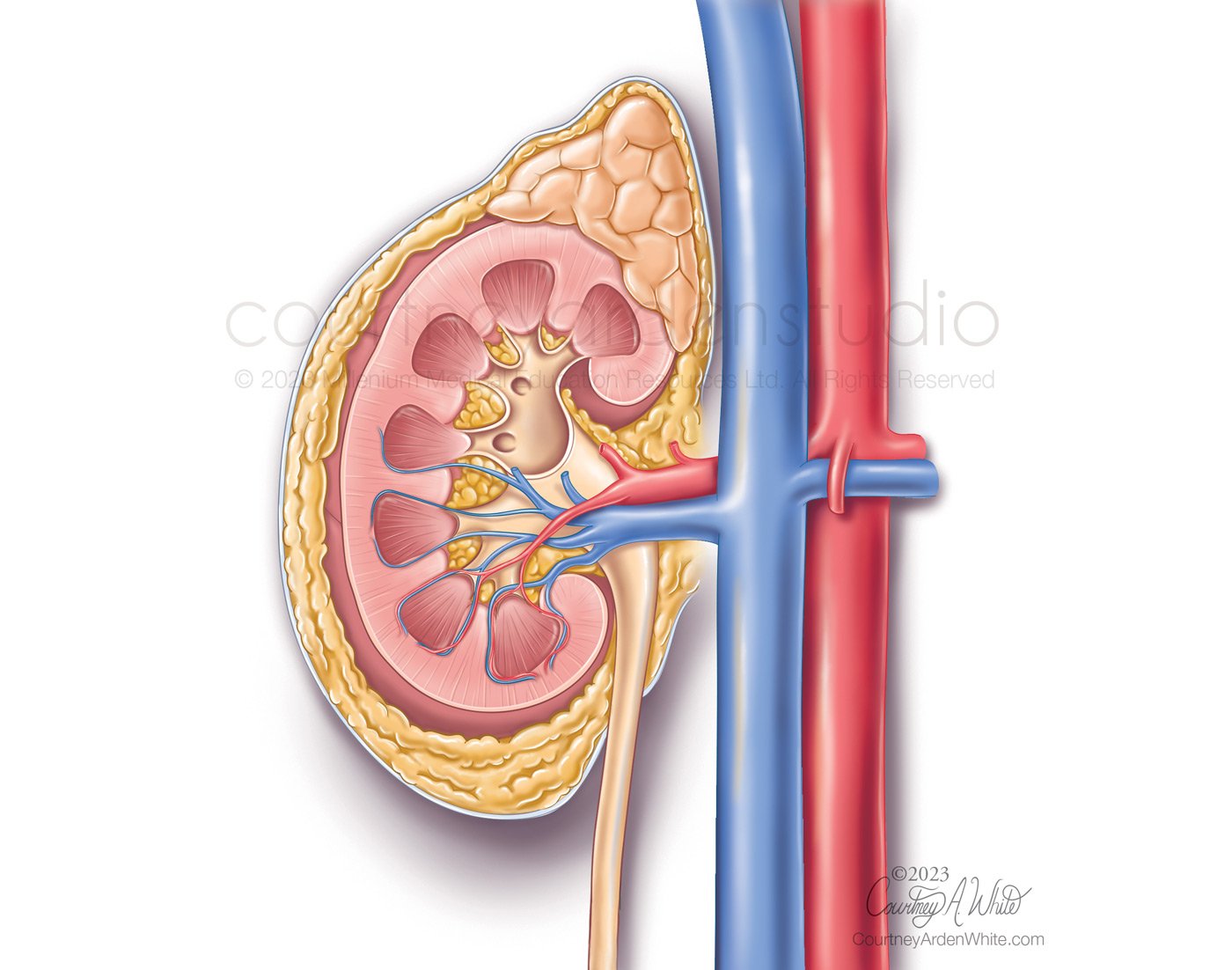
Urology, Reproduction and Gynecology Illustration: kidney cross-section anatomy
Urology, Reproduction and Gynecology Illustration: kidney cross-section
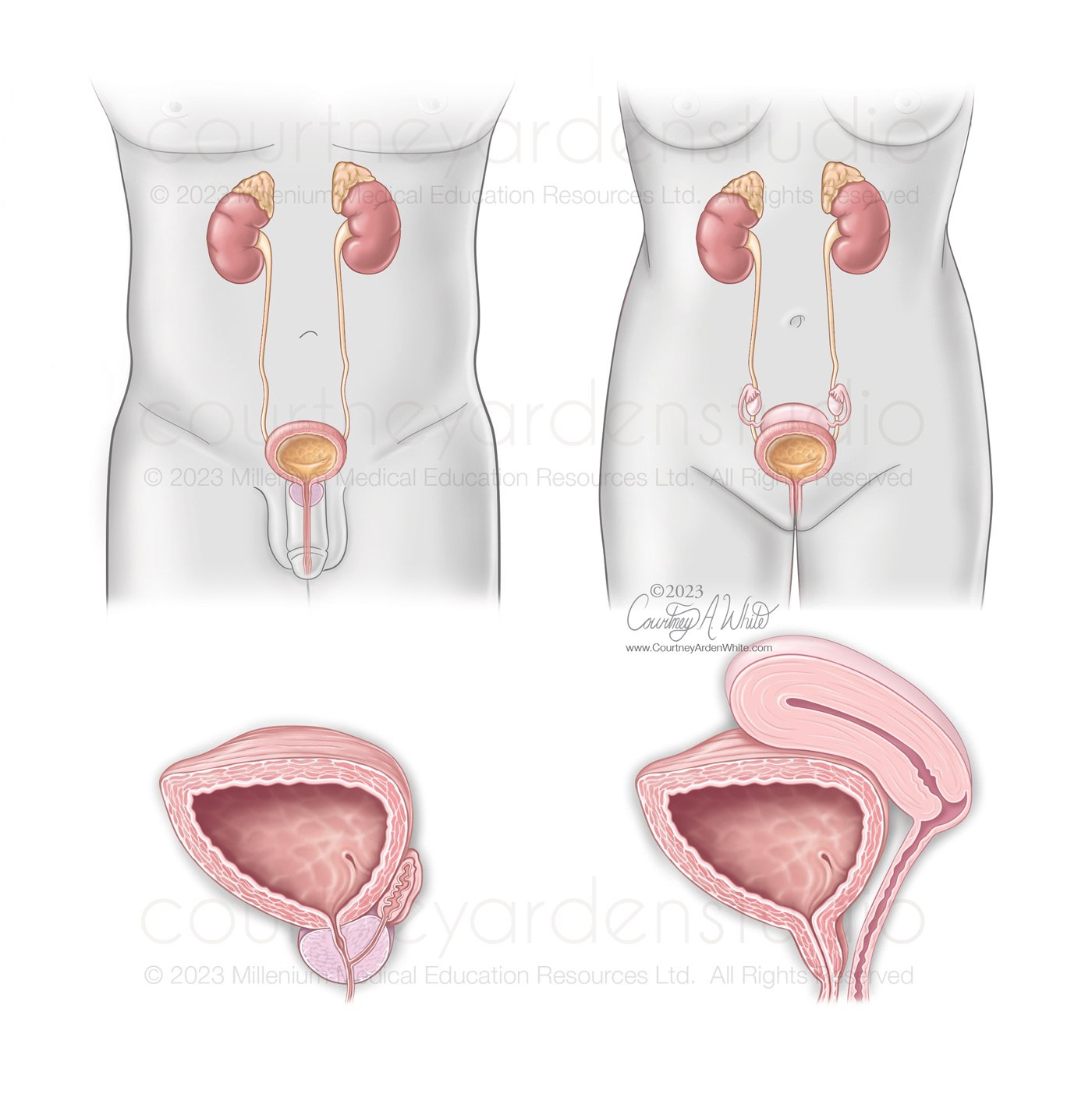
Urology, Reproduction and Gynecology Illustration: Female vs. Male anatomy of the the Urinary Tract and Reproductive Organs