
Cancer Illustration
This portfolio about Cancer Illustration shows cancer growth at all stages in many different parts of the body, including the kidneys, thyroid, prostate, bladder, breast, and liver.
Many of these illustrations were created for patient education or medical education for medical students.
Click on each illustration below to enlarge.

Cancer Illustration: About the Thyroid Gland: This is a patient education piece about the thyroid gland and thyroid cancer. It included a tent card and a double-sided informational sheet.
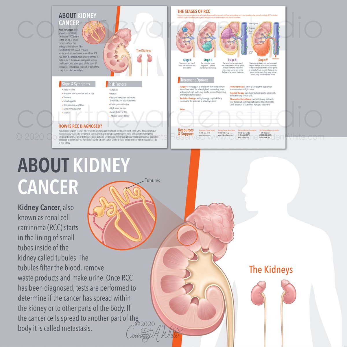
Cancer Illustration:Tear pad about kidney cancer

Cancer Illustration: Kidney Tumor

Cancer Illustration: The Stages of RCC

Cancer Illustration: Advanced prostate cancer. This illustration was created for a two-page patient education piece I wrote on advanced prostate cancer. When prostate cancer spreads beyond the prostate to nearby tissue, such as the seminal vesicles, bladder, or rectum, it's considered locally advanced (stage 3). If it spreads to other parts of the body, such as the lymph nodes, bones, liver, or lungs, it is considered advanced prostate cancer. By using visual aids like medical illustrations, healthcare providers can communicate important information to patients clearly and engagingly. I used bold, black outlines to align with the client's existing material on prostate cancer.

Cancer Illustration: Metastatic prostate cancer

Cancer Illustration: Chemotherapy and Affected Cells. This illustration was created for medical education on cancer treatment. Chemotherapy kills cells as they divide. Since cancer cells divide and create new cells more quickly, chemo will kill the cancer cells and spare the other cells in your body. However, specific cells in the body also divide and create new cells quickly, including blood cells in bone marrow, cells lining the GI tract, and cells of the hair follicles. Chemo can damage these healthy cells, causing side effects such as hair loss, anemia, infection, GI problems, and mouth, tongue, and throat problems.

Cancer Illustration: Cell Injury and Response to Hypoxemia

Cancer Illustration: Chemotherapy Administered-Cancer Returns Over time
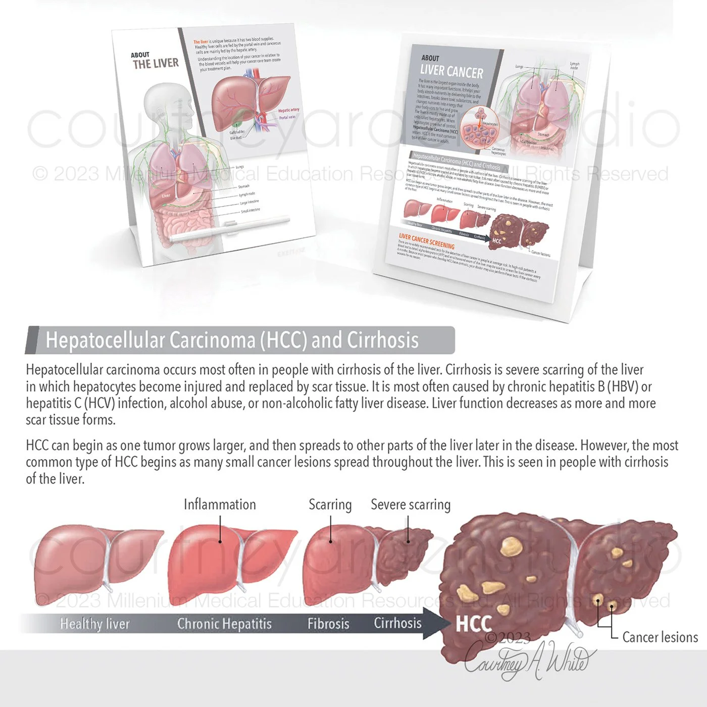
Cancer Illustration: Tear pad and tent card about hepatocellular carcinoma (HCC): These illustrations were made for a patient education tear pad and tent card about the liver and liver cancer. I designed and wrote the content for this piece as well. Hepatocellular carcinoma (HCC) is the most prevalent type of liver cancer in adults, and it typically occurs in people with cirrhosis of the liver. HCC usually starts as several small cancerous lesions that spread throughout the liver.

Cancer Illustration: Hepatocellular carcinoma (HCC) showing many small cancer lesions

Cancer Illustration: Myelofibrosis progression. This illustration was created for a patient education guide and folio on myelofibrosis. Myelofibrosis is a rare blood cancer that involves the buildup of scar tissue in the bone marrow, which interferes with the healthy production of blood cells. Because less red blood cells are made, it can lead to anemia.

Cancer Illustration: von Hippel-Lindau disease (VHL): This illustration was part of a patient education booklet about the kidneys and kidney diseases. It explains von Hippel-Lindau (VHL) disease, which is a rare condition that causes tumors to grow in different parts of the body. This disease is caused by a mutation in the VHL gene, and individuals with this gene mutation are at a higher risk of developing renal cell carcinoma (RCC).

Cancer Illustration: von Hippel-Lindau disease (VHL): This illustration was part of a patient education booklet about the kidneys and kidney diseases. It shows von Hippel-Lindau (VHL) disease, which is a rare condition that causes tumors to grow in different parts of the body.

Cancer Illustration: Kidney Cancer Cells in the Renal Tubules. This illustration was part of a patient education booklet about the kidneys and kidney diseases. This illustration shows how RCC starts in the cells that line the renal tubules.

Cancer Illustration: Fuhrman grade kidney cancer cells. This illustration was part of a patient education booklet about the kidneys and kidney diseases. It depicts the Fuhrman grade scale, which is used to describe the aggressiveness of RCC cells and ranges from grade 1 to grade 4.

Cancer Illustration: kidney cancer booklet cover

Cancer Illustration: kidney cancer staging T3 illustration
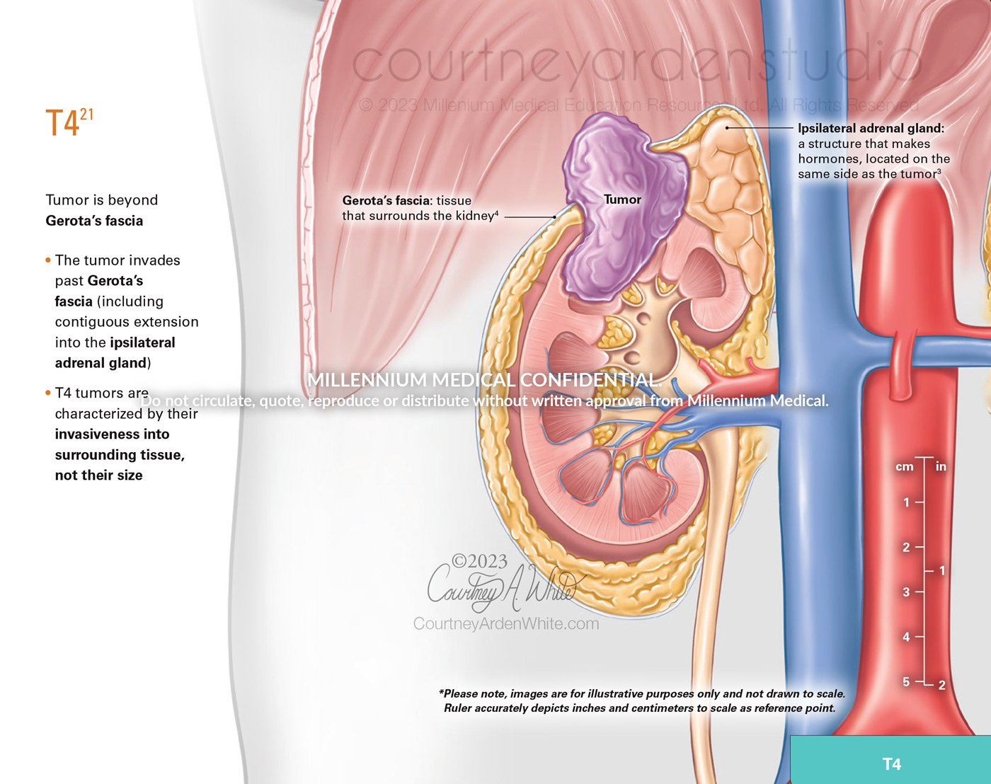
Cancer Illustration: kidney cancer staging T4 illustration

Cancer Illustration: kidney cancer booklet about the kidneys page

Cancer Illustration: Common Features of TNBC. This illustration was created for a two-page patient education tear pad on metastatic triple-negative breast cancer (mTNBC). The term triple-negative refers to three receptors commonly found in breast cancer: the estrogen receptor (ER), progesterone receptor (PR), and human epidermal growth factor receptor 2 (HER2). If the cancer cells don't express these, it is called triple negative.

Cancer Illustration: Illustrated Microscopy of BM invasion

Cancer Illustration: Bladder Health & Bladder Cancer Flip Book Cover

Cancer Illustration: Bladder Health & Bladder Cancer Flip Book - What is Bladder Cancer?

Cancer Illustration: Bladder Health & Bladder Cancer Flip Book - Papillary carcinoma vs Carcinoma in situ (CIS) - Papillary grow in thin, finger-like projections. CIS is a flat tumor that does not grow toward the hollow part of the bladder and is only in the inner layer of the bladder wall.
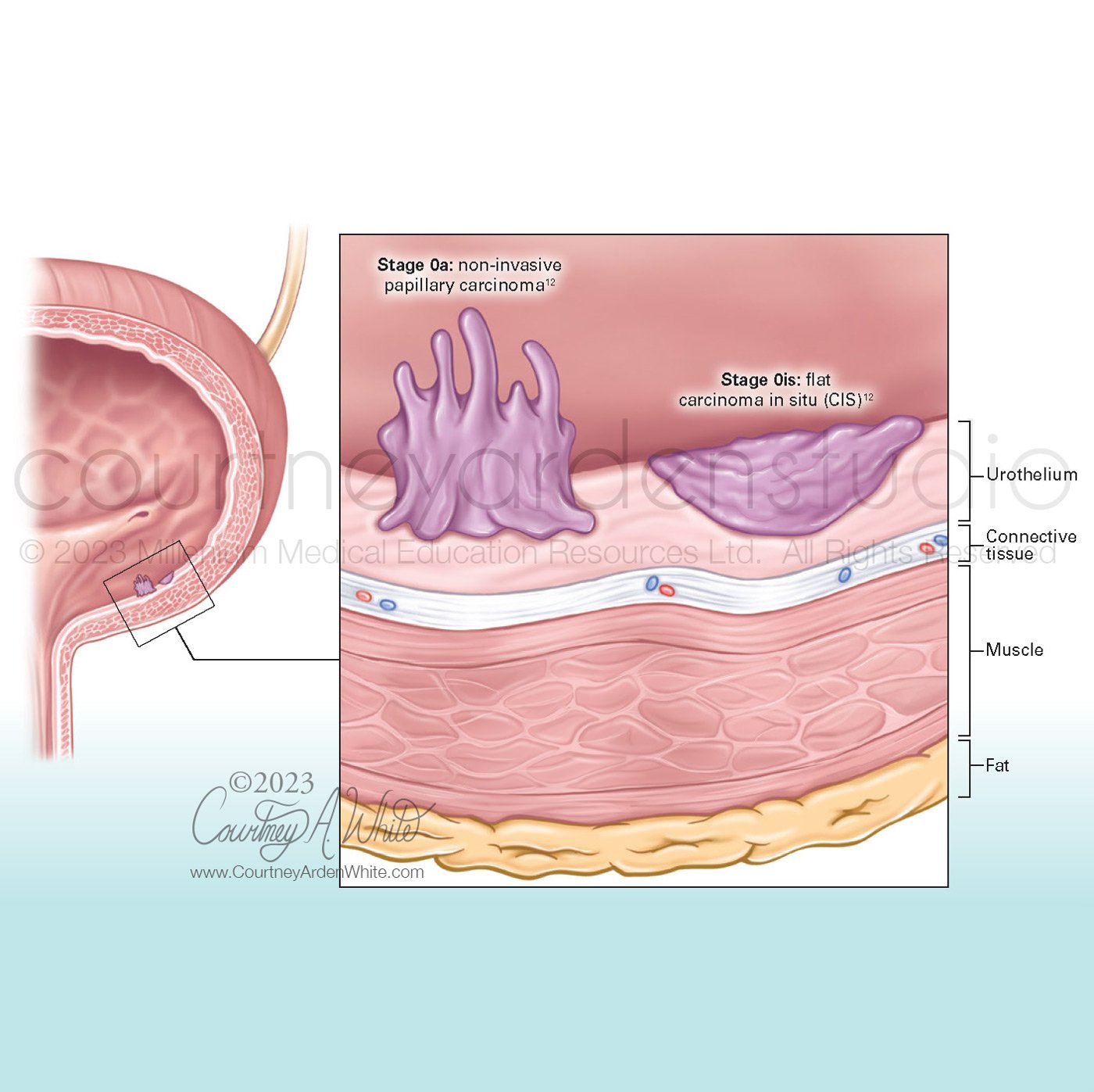
Cancer Illustration: Bladder Health & Bladder Cancer Flip Book - Stage 0

Cancer Illustration: Bladder Health & Bladder Cancer Flip Book - Stage 2

Cancer Illustration: Bladder Health & Bladder Cancer Flip Book - Stage 3

Cancer Illustration: Bladder Health & Bladder Cancer Flip Book - Stage 4

Cancer Illustration: Bladder Health & Bladder Cancer Flip Book - Low grade vs. high grade cells