
The Digestive System Illustrations
The digestive system illustrations below include anatomy illustrations of the stomach, intestines, esophagus, pancreas, oral cavity, nasal cavity, and pharynx.
These illustrations also cover the mechanisms of swallowing, as well as medical conditions such as celiac disease, herniation and intussusception. Additionally, there are cellular illustrations displaying the layers of the digestive tract and details on absorption.
Click on each image below to enlarge.

Digestive System Illustrations: Anatomy of the Palate, Nasal Septum and Pharynx

Digestive System Illustrations: Orientation of the Oral Cavity, Nasal Cavity and Pharynx

Digestive System Illustrations: Phases of Swallowing

Digestive System Illustrations: Actions of Pharyngeal Phase

Digestive System Illustrations: Colon Vasculature - This illustration depicts the vasculature of the colon. As there are many terms used in this illustration, we have color-coded the different branches to simplify the learning process for medical students. The color-coding helps students to understand which branches supply each part of the colon.

Digestive System Illustrations: Motion of the Colon: Mass Movement - This illustration explains the fast movements of the colon called mass movements. The colon contracts and relaxes, which helps to move the chyme through the large intestine until it reaches the rectum. During this process, the chyme absorbs water and electrolytes. This happens around three times a day. If the frequency of movement increases, it can cause diarrhea; if it decreases, it can cause constipation.

Digestive System Illustrations: Segmentation and Peristalsis - This illustration about segmentation and peristalsis of the small intestine was created for medical education. Segmentation and peristalsis are two ways the muscles of the small intestine moves the contents or chyme through the tract. Segmentation causes a mixing motion to allow for absorption of water and nutrients. Peristalsis causes the chyme to propel forward along the tract.

Digestive System Illustrations: Relative Anatomy of the Stomach - This illustration was created for medical education for medical students about the relative anatomy of the stomach. The stomach is located in the upper left quadrant of the abdomen. It is partially covered anteriorly by the liver and is located anterior to the pancreas and left kidney. The lesser curvature of the stomach is connected to the liver by the lesser omentum, and the greater curvature of the stomach is connected to the greater omentum, which drapes over the intestines.

Digestive System Illustrations: How Pancreatitis Happens
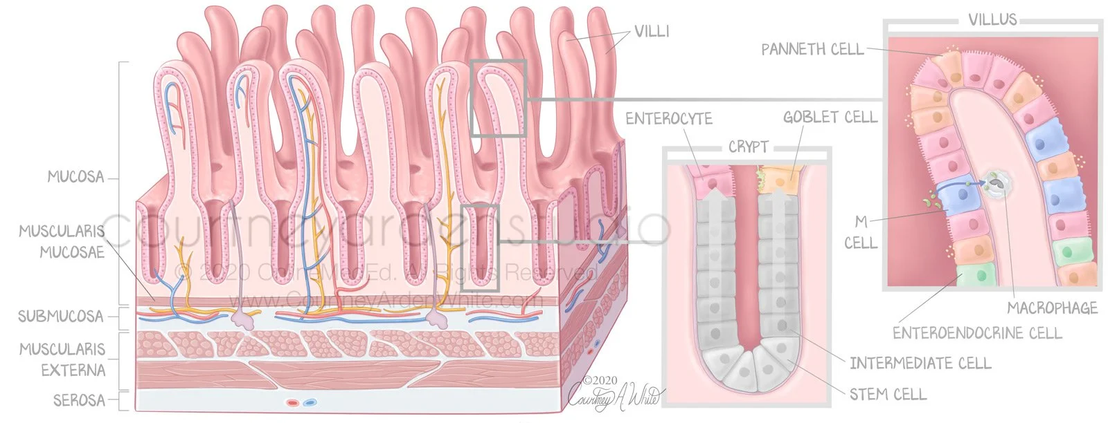
Digestive System Illustrations: Crypts, Villi, and Cells

Digestive System Illustrations: Regional Differences of Mucosa
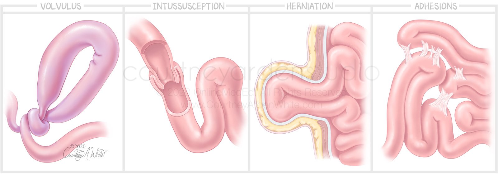
Digestive System Illustrations: Causes of Bowel Obstruction

Osmotic Diarrhea

Digestive System Illustrations: Site of Absorption - This illustration about the GI tract was created for medical education. It shows where different nutrients are absorbed in the digestive tract. Most absorption occurs in the small intestine, which consists of three parts: the duodenum, the jejunum, and the ileum. After the ileum comes the large intestine, where any remaining bile acids that were not absorbed in the ileum are absorbed.

Digestive System Illustrations: General Histology of the Stomach

Digestive System Illustrations: Fat Absorption

Digestive System Illustrations: Absorption Summary: Proteins, Fats and Carbs
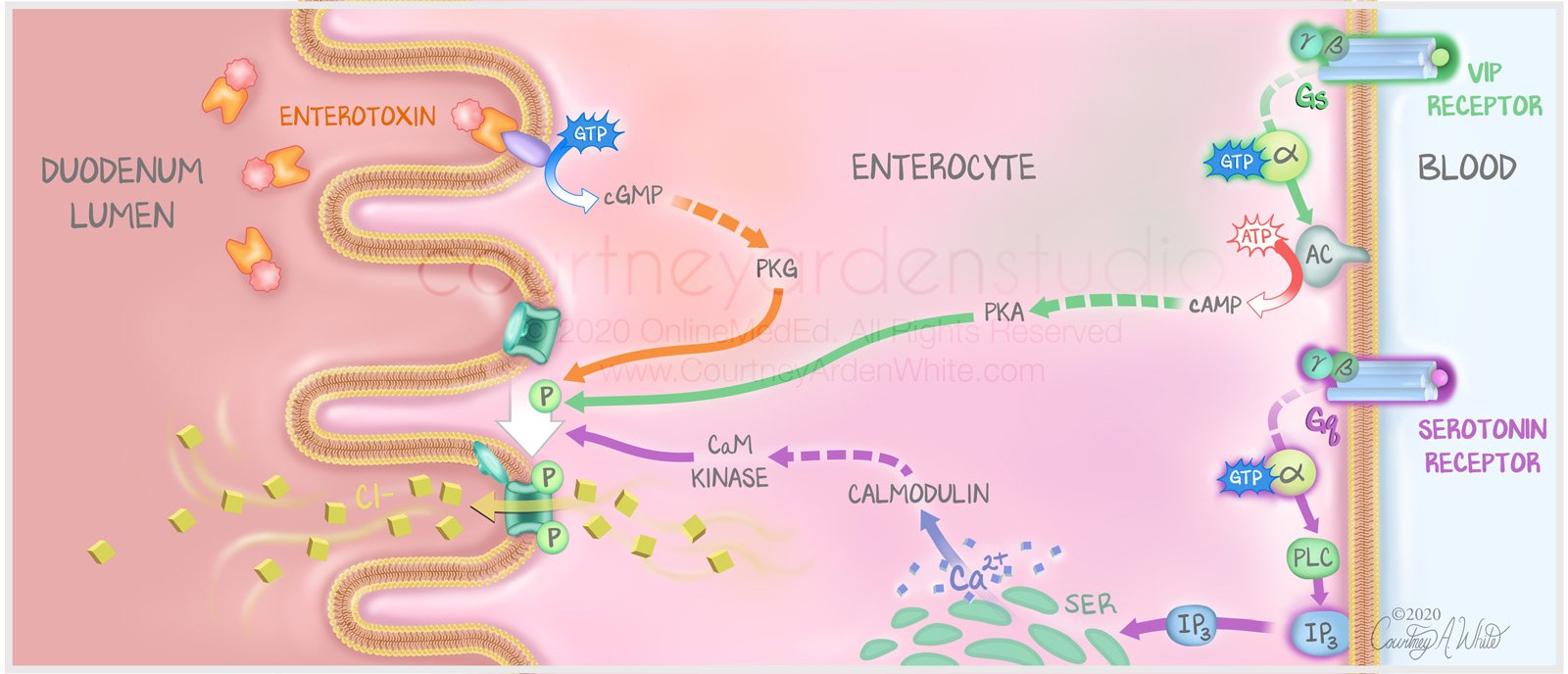
Digestive System Illustrations: Mechanisms of Secretory Diarrhea

Digestive System Illustrations: Hernias

Digestive System Illustrations: Severity of Hernias

Digestive System Illustrations: Celiac Histology

Digestive System Illustrations: Crohn's vs Ulcerative Colitis - This medical illustration was designed for medical educational purposes to highlight the differences between ulcerative colitis and Crohn's disease, both of which are inflammatory bowel diseases (IBD). While Crohn's can cause inflammation anywhere in the gastrointestinal tract, including the mouth, ulcerative colitis only causes inflammation and ulceration in the large intestine. Additionally, ulcerative colitis affects only the inner layer of the large intestine wall, whereas Crohn's can reach all layers, as shown in the illustration by the knife-like fissures. Furthermore, ulcerative colitis causes continuous lesions or inflammation, while Crohn's has skip lesions where there will be a healthy part of the intestine between two inflamed parts.

Digestive System Illustrations: Celiac Disease

Digestive System Illustrations: Megacolon
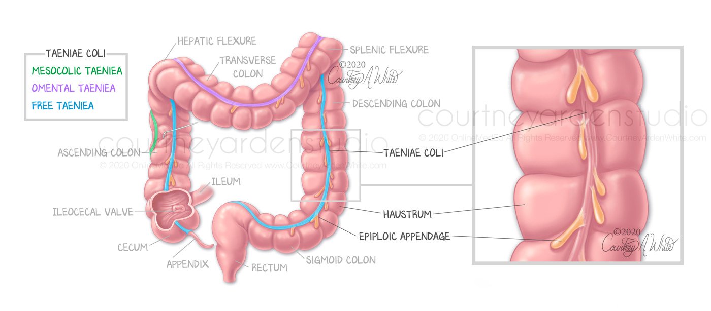
Digestive System Illustrations: Anatomy of the Colon
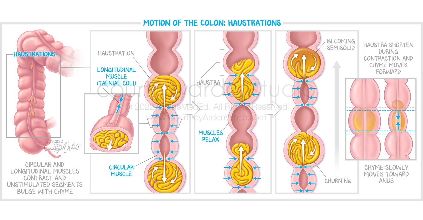
Digestive System Illustrations: Motion of the Colon: Haustrations
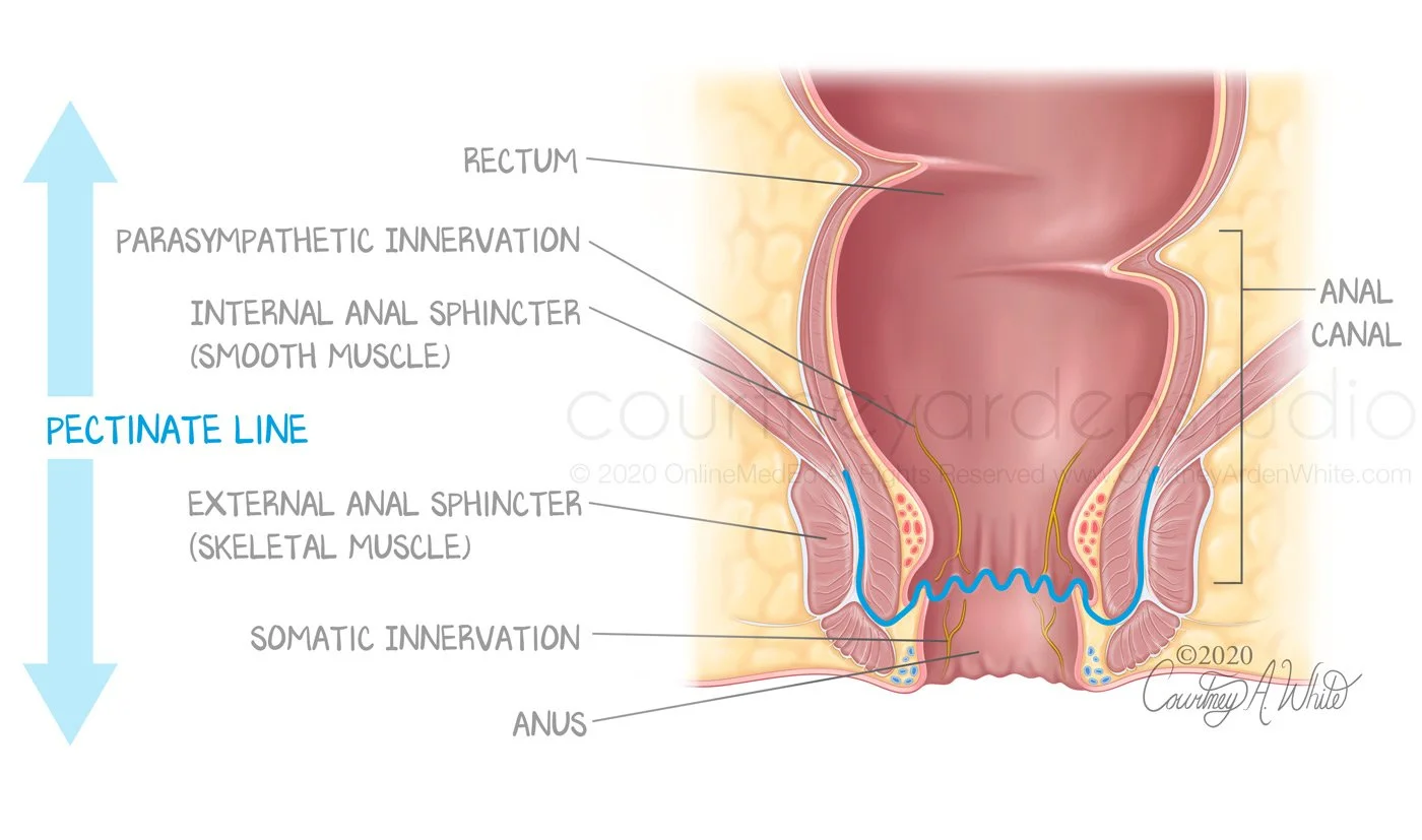
Digestive System Illustrations: Pectinate Line